Anatomy, Cerebrum vs Cerebellum, Brain Stem, Subcortical Cortex, Cerebral Cortex [MCAT, USMLE, Biology, Medicine] – Moosmosis
[ad_1]
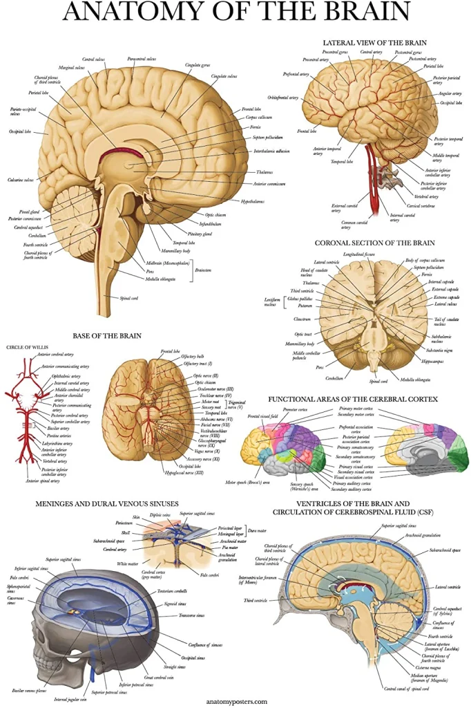
In this lesson, we explore the nervous system and share notes as part of the study guide series. We will explore the awesome brain and nerves! Topics include the nervous system anatomy, cerebellum, brain stem, subcortical cortex, cerebral cortex, and cerebrum.
Check out our popular nervous system notes.
Cerebellum
- Recall, the cerebellum is behind the brainstem, underneath the cerebrum. It is also divided into left and right hemispheres, and has many different functions.
- Cerebellum is most notable for coordinating movement; it smooths moves out & increases accuracy.
- Three parts to how information travels into and out of the cerebellum to let it coordinate movement:
- Motor Plan — this involves which muscles need to contract and at what intensity and duration.
- While the movement is actually being executed by UMN through LMN, information about the motor plan is delivered from the cerebrum to the cerebellum, as well.
- Position Sense — Muscle spindles, e.g. will send info through somatosensory neurons. Once it enters brain stem and cerebellum, the latter can tell if it’s going according to plan or if corrections are necessary. Usually that is the case, it needs some sort of correction to make the movement match the motor plan, so the cerebellum needs to send feedback..
- Feedback — After receiving info about the position sense, the cerebellum may send feedback back to the motor areas of the cerebrum, the areas that came up with the motor plan in the first place, to try and correct the movement while it’s occurring by changing activity of the UMN.
- Motor Plan — this involves which muscles need to contract and at what intensity and duration.
- Note: cerebellum is set up such that the middle of the cerebellum tends to coordinate movement of the middle body, most notably walking. The part of the cerebellum more on the side is more involved in movement of the limbs. Many parts of the cerebellum also coordinate movements of speech and of our eyes.
Brainstem
- Brainstem basically connects all parts of the nervous system: the cerebrum, cerebellum, and spinal cord. It also connects all the cranial nerves.
- Inside of the brain stem has some similarities to the spinal cord, particularly in the medulla. Most of the white matter is on the outside and most of the gray matter is on the inside, but it’s more mixed than in the spinal cord.
- Much of the brain stem gray matter are sort of distributed or scattered neurons not in distinct groups or bundles. This is the reticular formation of the brain stem. It plays an important role in many autonomic functions, such as circulation, respiration, and digestion.
- Reticular formation also sends lots of axons up to the cerebrum and it plays a role in some of the higher functions, as well, including cognition, emotion, and consciousness.
- A lot of the white matter passing through the brain stem is actually connecting different parts of the nervous system. Long tracts are collections of axons that often connect the cerebrum to the spinal cord. Two big categories of long tracts to which the brain stem plays host:
- Upper Motor Neuons (important for movement)
- Most of the 12 pairs of cranial nerves humans have are attached to the brain stem.
- These nerves perform motor functions, sensory functions (many different kinds of sense functions), and a number of automatic functions.
- These nerves are related to a lot of the gray matter inside the brain stem. In addition to the reticular formation, there are collections of neuron somas that are distinct nuclei. Cranial nerves often carry info into or away from these nuclei. — ex: nuclei have neuron somas and axons leave the brain stem through cranial nerves to perform motor functions.
- Cranial nerves mostly perform their functions in the head and neck, but there are a few that travel down the brainstem all the way to the body and perform functions in the trunk and limbs
- Ex of cranial nerve functions: sensation of the face, movements of the eyes / face / jaw / throat, influence the heart and intestines.

Subcortical Cerebrum
- This is the deep part of the cerebrum, under the cortex of gray matter. Deep white
matter and deep gray matter (which are called nuclei) are subcortical - Lots of white matter deep in the cerebrum, which contains axons going from cerebral
cortex gray matter and/or from deeper nuclei and/or to and from the brainstem - Internal capsule (pink) – subcortical band of white matter, shaped like a V (if looking
top-down). Contains corticospinal tract of UMN. - Basal ganglia (blue) — collection of subcortical nuclei that function
together; play a major role in motor functions. (They don’t contain UMN
themselves but help out the motor areas of cerebral cortex). Also
contributes to cognition and emotion. - Corpus callosum (purple) — band of white matter connecting left and
right hemisphere, allows info to travel across them. - Thalamus (Diencephalon) (yellow) — Under the corpus callosum.
Plays an important role in sensory functions. Almost all the senses have
pathways that travel to the thalamus for sensory processing and then
travel further on to areas of the cerebral cortex. - Thalamus is also very important for all higher functions of the brain (cognition, emotion, and consciousness) bc it is connected to many brain areas and plays a role in passing info around between them and other areas / subcortical structures.
- Hypothalamus (green) — Under the thalamus. It is connected to and controls the pituitary gland (circled in green), aka “the master gland” that links nervous and endocrine systems and plays a major role in controlling glands. The hypothalamus is also involved in higher functions.

Cerebral Cortex
- Cerebral cortex is layer of gray matter on outside of the cerebrum. It has many ridges called gyri (sing: gyrus), and small grooves on either side of a gyrus called sulci (sing: sulcus).
- Large grooves separating lobes are called fissures.
- Cerebral cortex is divided into lobes, named for the bones of the
skull right above them. - Frontal — logic and decision making
- Parietal — proprioception and sensory
- Temporal — language and memory, olfactory, auditory
- Occipital — vision
- A few senses and motor functions of cerebral cortex on one side of
the brain tend to be involved with the other side of the body. - Visual information coming in on the right side of a body will be
processed on the left side of the brain (in the occipital lobe),
and vice versa - Somatosensory information, such as a hot or cold something applied to the skin on the right side, will end up being processed and brought to consciousness in the parietal cortex
- Motor functions for, e.g., the right leg will be processed on the left side of the brain (specifically, the back part of the frontal lobe).
- Other senses (besides vision and somatosensation) tend to get processed in areas of the cerebral cortex on both sides
- We can divide the areas of the cerebral cortex based on the function of that area:
- Primary cortex: performs basic motor or sensory functions
- Association cortex: associates different types of info to do more complex processing and functions.
- ex: For areas of motor cortex, there’s primary motor cortex/cortices that do basic motor functions, and then association motor cortices do more complex functions like planning of movements. Some areas of association motor cortices take in different types of information and integrate / process it to do higher level complex motor or sensory functions, and to produce higher functions of the nervous system such as cognition and emotion.
- One aspect of cognition is language (ability to turn thoughts into words), performed by certain areas of cerebral cortex in left hemisphere.
- Cerebral cortex on both sides plays a role in attention but, for most people, the right cerebral hemisphere’s cortex plays a role in paying attention to both sides of the body and the environment. (The left hemisphere just seems to pay more attention to the right side of the body).

Support Us at Moosmosis.org!
Thank you for visiting, and we hope you find our free content helpful! Our site is run 100% by volunteers from around the world. Please help support us by buying us a warm cup of coffee! Many thanks to the kind and generous supporters and donors for doing so! 🙂
Copyright © 2022 Moosmosis Organization: All Rights Reserved
All rights reserved. This essay first published on moosmosis.org or any portion thereof may not be reproduced or used in any manner whatsoever
without the express written permission of the publisher at moosmosis.org.

Please Like and Subscribe to our Email List at moosmosis.org, Facebook, Twitter, Youtube to support our open-access youth education initiatives! 🙂
[ad_2]
Source link

